Phospholipases PLBD1 and PLBD2: Difference between revisions
Tomemerald (talk | contribs) |
Tomemerald (talk | contribs) |
||
| Line 87: | Line 87: | ||
While this merely compares human PLBD1 and PLBD2, the collected reference sequences (intronated against their respective genome assemblies) confirm that introns in both genes are deeply conserved. | While this merely compares human PLBD1 and PLBD2, the collected reference sequences (intronated against their respective genome assemblies) confirm that introns in both genes are deeply conserved. | ||
PLBD1 introns do not correspond well to those of PLBD2: | |||
>PLBD1_homSap Homo sapiens (human) first and last introns are not mappable | >PLBD1_homSap Homo sapiens (human) first and last introns are not mappable | ||
0 MTRGGPGGRPGLPQPPPLLLLLLLLPLLLVTAEPPKPA 1 | 0 MTRGGPGGRPGLPQPPPLLLLLLLLPLLLVTAEPPKPA 1 | ||
Revision as of 19:44, 27 October 2010
Introduction
A surprising number of orphan human enzymes (unknown substrate) still exist ten years after the completion of the human genome. PLBD1 and PLBD2 are semi-orphans in the sense of being probable phospholipases of B class but with uncertain physiological substrates and thus functionalities. This is especially important in the case of PLBD2 which localizes to the lysosome, as its absence could plausibly lead to a serious yet unrecognized lysosomal storage disease.
No bioinformatic algorithm or experimental protocol leads with any certainty to determination of function. The gene pair here has seven targeted publications but cases exist where protein function remains unknown after ten thousand papers (eg PRNP).
PLBD1 and PLBD2 constiture a small gene family (sequence homology class) within vertebrates though it occurs expanded in some early eukaryotes. However, the Pfam clan (NTN: N-terminal nucleophile aminohydrolases) may have additional representatives in humans diverged beyond recognizability in primary sequence among its ten family members. These establish the great antiquity of the fold and certain of its features but are not likely to shed additional light on phospholipases.
PLBD2 presents a special difficulty in that a sequence of post-translational steps are necessary for its activation. Without these, potential substrates can hardly be assayed. These steps include removal of the signal peptide, mannosylation appropriate to the lysosome targeting receptor, and self-catalytic proteolytic activation (into 28k and 42k fragments which remain associated) to expose the substrate binding site once this is appropriate.
Because PLBD1 and PLBD2 are full length paralogs, the bioinformatic approach below considers both. PLBD1 has been somewhat more amenable to activation whereas PLBD2 has a high-resolution structual determination. Thus comparative genomics allow for annotation transfer, first from PLBD2 to a structural model for PLBD1 (provided by SwissModel), then transfer of PLBD1 experimental successes to PLBD2.
However the gene duplication event occured some 650 million years ago and the two genes are quite diverged today. Yet certain core features remain conserved, including the fold, active site residues, signature motifs, ertain glycosylation sites and even the fragmentation pattern, implying these are essential functional features under long-range strong selective pressure for their maintainence.
The disulfides are only separately conserved but this fortuitously provides a reliable signature for assigning deeply diverged proteins from early eukaryotes to their ortholgy class. As the respective functions become known, we can hope to better understand how the gene duplication event contributed advantageously to increasing evolutionary complexity, requiring both copies to persist in most species over the immense time spans involved.
Conservation at critical sites
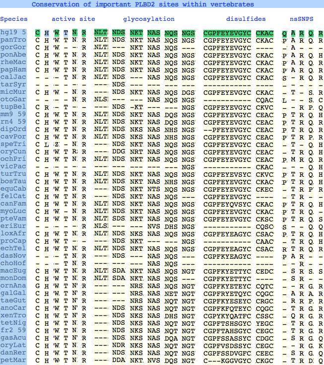
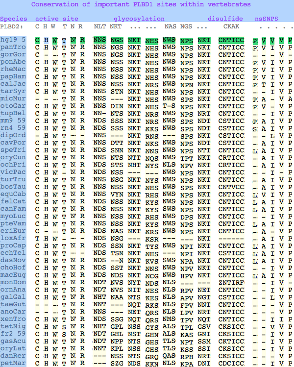
The six residues of PLBD2 associated with the active site are completely conserved within vertebrates to within genomic sequencing error. These same six residues are also completely conserved within PLBD1. Indeed 3 of the residues are conserved in the broader NTN hydrolase clan.
This is perhaps unsurprising since the active site was established a couple billion years earlier in the bacterial ancestor. However if PLBD2 and PLBD1 have different substrates, this establishes that these six residues are insufficient to distinguish the two active sites. Note H266 and T330 do not contribute their side chain, leaving them and W269 to separate phospholipases from the other NTN hydrolases.
The glycosylation sites are surprisingly conserved both within and between PLBD2 and PLBD1. Some of the motifs may be either recently acquired within later vertebrates or spurious glycosylation motifs with N and D both acceptable (or similar small amino acids) in the first slot of the NxS/T motif. Glycosylation is important in correct targeting of lysosomal proteins, more so than in generic endoplasmic reticulum proteins where motifs are often poorly conserved (as in sulfatases).
PLBD2 has two established disulfides. Strict sequence conservation of these throughout vertebrates (indeed, throughout metazoa) suggests both play an important role in protein structure and stability.
In PLBD1 however, the first disulfide is not a possibility and while an opportunity exists for a disulfide homologous to the the second disulfide of PLBD2, indels cloud the alignment and spacing would have to be different. There is additionally ambiguity given C...CC as to the cysteines involved. Indeed a second distal disulfide may occur utilizing C...CC.............C which has no counterpart in PLBD2. While cysteines can be conserved for many reasons other than disulfide (as in the nucleophile cysteine here), suitably proximity and side chain orientation in the SwissModel of PLBD1 would argue for disulfide. Comparative genomics suggests that C2 and C4 may form an ancient disulfide whereas C1 and C3 might represent a deuterostome innovation.
homSap CNTICCREDLNSPNPSPGGC human PLBD1
braFlo CSAICCRKDLAKVGAKPDGC Branchiostoma floridae
strPur SKSICMRGDLM-TSPMPNGC Strongylocentrotus purpuratus XM_001192029
nemVec MNAICSRGDLIADGPRASGC Nematostella vectensis XM_001638165
monBre YNAICSRGDLESDSPSPGGC Monosiga brevicollis XM_001745398

SwissModel coordinates for PLBD1 show the 2nd and 4th sulfur atoms separated by 2.03 angstroms:
ATOM   3552  SG  CYS   471      49.680 -13.769 -12.461
ATOM   3579  SG  CYS   475      49.273 -14.310  -4.881
ATOM   3585  SG  CYS   476      51.067  -9.716  -9.172
ATOM   3678  SG  CYS   490      50.737 -13.198  -5.75
The known human SNPs of PLBD2 are in some cases quite radical substitutions in terms of both physical qualities of the substituted amino acid and the degree of observed phylogenetic conservation at that site. These likely result in unstable and/or inactive enzyme. Both enzymes are autosomal so compensation might occur in the recessive state, or alternately, PLBD2 and PLBD1 could fill for each other to some extent. In either case, lysosomal storage disease might not be clinically observable.
Here Q54P may actually be a mutation in the reference sequence individual (with the SNP representing wildtype) as proline is quite well conserved throughout mammals. In A204V, valine is quite a bulky substituent for a site normally restricted to small amino acids; R354C is definitely a serious mutation, no doubt attributable to a CpG hotspot; Q521K appears milder as does R524C.
The known human SNPs of PLBD1 can be analyzed similarlly. P26Q and V30L may be inconsequential as they occur in the rather unconstrained primary sequence of the N-terminus; V265I occurs at an ILV reduced alphabet; V377A and P534A are much more serious despite the aliphatic nature of alanine and likely give rise to dysfunctional protein.
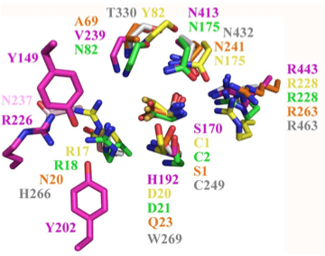
Structural superposition of active sites from five NTN hydrolases
showing conserved side chains (*) and relevant main chains (....)
(adapted from Fig 6 of Lakomek et al. BMC Struct Biol.2009;9:56:)
                                            *         (*)       *    *
PLBD2 phospholipase B-like     gray   3FGR  C244 H261 W264 T325 N427 R458 human numbering
PLBD1 phospholipase B-like     ....   pred  C228 H245 W248 T303 N402 R433 human numbering SwissModel
Cephalosporin acylase          pink   1OQZ  S170 .... H192 .... N413 R443
Conjugated bile acid hydrolase green  2BJF  C2   .... D21  .... N175 R228
Penicillin V acylase           yellow 3PVA  C1   .... D20  .... N175 R228
Penicillin G acylase           orange 1K5S  S1   .... Q23  .... N241 R263

Human SNPs resulting in amino acid substitions:
PLBD2:                PLBD1:
 Q54P   rs7965471    P26Q   rs1141509
 A204V  rs12231990   V30L   rs12296104
 R354C  rs56935204   V265I  rs7957558
 Q521K  rs17852787   V377A  rs2287541
 R524C  rs12425042   P534A  rs1600
Intron evolution
PLBD1 and PLBD2, being full length paralogs, clearly indicate an early gene duplication and subsequent divergence to the current low percent identity. Segmental duplications preserve any introns present at the time of the event and these generally persist in both position and phase into living species.
However PLBD1 and PLBD2 -- despite having similar numbers of introns -- exhibit very little in common in terms of location as the diagram below shows. One possibility is that a second copy arose as a retroprocessed gene (a mechanism erasing existing introns) and was subsequently intronated at random positions. This is unlikely here given that 10-11 relatively rare events would be needed.
The remaining possiblity is that the gene duplication took place prior to the main era in early eukaryotes during which the bulk of introns were established. This fits the current state of high divergence despite fairly slow rates of evolution during metazoan times.
The last five amino acids of each PLBD1 exon are colored below. Then using an alignment of PLBD1 to PLBD2, the colors are mapped to the homologous five residues within PLBD2. There they fall on the ends of exons only when these correspond to those of PLBD1. The outcome here -- despite uncertainties in alignment gapping -- shows intron positions do not correspond with the exception of the terminal intron (which also is phase 0).
While this merely compares human PLBD1 and PLBD2, the collected reference sequences (intronated against their respective genome assemblies) confirm that introns in both genes are deeply conserved.
PLBD1 introns do not correspond well to those of PLBD2:

>PLBD1_homSap Homo sapiens (human) first and last introns are not mappable
0 MTRGGPGGRPGLPQPPPLLLLLLLLPLLLVTAEPPKPA 1
2 GVYYATAYWMPAEKTVQVKNVMDKNGDAYGFYNNSVKTTGWGILEIRAGYGSQTLSNEIIMFVAGFLEGYLTAP 2
1 HMNDHYTNLYPQLITKPSIMDKVQDFME 2
1 KQDKWTRKNIKEYKTDSFWRHTGYVMAQIDGLYVGAKKRAILEGTK 0
0 PMTLFQIQFLNSVGDLLDLIPSLSPTKNGSLKVFKRWDMGHCSALIK 0
0 VLPGFENILFAHSSWYTYAAMLRIYKHWDFNVIDKDTSSSRLSFSSYP 1
2 GFLESLDDFYILSSGLILLQTTNSVFNKTLLKQVIPETLLSWQRVRVANMMADSGKRWADIFSKYNS 1
2 GTYNNQYMVLDLKKVKLNHSLDKGTLYIVEQIPTYVEYSEQTDVLRK 1
2 GYWPSYNVPFHEKIYNWSGYPLLVQKLGLDYSYDLAPRAKIFRRDQGKVTDTASMKYIMRYN 1
2 NYKKDPYSRGDPCNTICCREDLNSPNPSPGGCYDTK 0
0 VADIYLASQYTSYAISGPTVQGGLPVFRWDRFNKTLHQGMPEVYNFDFITMKPILKLDIK* 0

>PLBD2_homSap Homo sapiens (human)
0 MVGQMYCYPGSHLARALTRALALALVLALLVGPFLSGLAGAIPAPGGRWARDGQVPPASRSRSVLLDVSAGQLLMVDGRHPDAVAWANLTNAIRETG 2
1 WAFLELGTSGQYNDSLQAYAAGVVEAAVSEE 0
0 LIYMHWMNTVVNYCGPFEYEVGYCERLKSFLEANLEWMQEEMESNPDSPYWHQ 0
0 VRLTLLQLKGLEDSYEGRVSFPAGKFTIKPLGFL 2
1 LLQLSGDLEDLELALNKTKIKPSLGSGSCSALIKLLPGQSDLLVAHNTWNNYQHMLRVIKKYWLQFREGPW 1
2 GDYPLVPGNKLVFSSYPGTIFSCDDFYILGSGL 0
0 VTLETTIGNKNPALWKYVRPRGCVLEWVRNIVANRLASDGATWADIFKRFNSGT 2
1 YNNQWMIVDYKAFIPGGPSPGSRVLTILEQIP 2
1 GMVVVADKTSELYQKTYWASYNIP 2
1 SFETVFNASGLQALVAQYGDWFSYDGSPRAQIFRRNQSLVQDMDSMVRLMR 2
1 YNDFLHDPLSLCKACNPQPNGENAISARSDLNPANGSYPFQALRQRSHGGIDVK 0
0 VTSMSLARILSLLAASGPTWDQVPPFQWSTSPFSGLLHMGQPDLWKFAPVKVSWD* 0
PLBD1 reference sequences
>PLBD1_homSap Homo sapiens (human) FLJ22662 PMID: 19019078,20093120
0 MTRGGPGGRPGLPQPPPLLLLLLLLPLLLVTAEPPKPA 1
2 GVYYATAYWMPAEKTVQVKNVMDKNGDAYGFYNNSVKTTGWGILEIRAGYGSQTLSNEIIMFVAGFLEGYLTAP 2
1 HMNDHYTNLYPQLITKPSIMDKVQDFME 2
1 KQDKWTRKNIKEYKTDSFWRHTGYVMAQIDGLYVGAKKRAILEGTK 0
0 PMTLFQIQFLNSVGDLLDLIPSLSPTKNGSLKVFKRWDMGHCSALIK 0
0 VLPGFENILFAHSSWYTYAAMLRIYKHWDFNVIDKDTSSSRLSFSSYP 1
2 GFLESLDDFYILSSGLILLQTTNSVFNKTLLKQVIPETLLSWQRVRVANMMADSGKRWADIFSKYNS 1
2 GTYNNQYMVLDLKKVKLNHSLDKGTLYIVEQIPTYVEYSEQTDVLRK 1
2 GYWPSYNVPFHEKIYNWSGYPLLVQKLGLDYSYDLAPRAKIFRRDQGKVTDTASMKYIMRYN 1
2 NYKKDPYSRGDPCNTICCREDLNSPNPSPGGCYDTK 0
0 VADIYLASQYTSYAISGPTVQGGLPVFRWDRFNKTLHQGMPEVYNFDFITMKPILKLDIK* 0

>PLBD1_braFlo Branchiostoma floridae (lancelet) XM_002595538
0 MEGRACRSCRLHHLSAVFLLFLVTIAA 1
2 GAEIQATAYLQAQGKVQVKLGVLDKQNGDAVATYDDR 2
1 LTENGWGVLNVVSGFGPKKLSDNDIMYLAGYLEGVLTQE 2
1 RIYQHYLNLYGIFFMGKSEDLVGK 0
0 VKKFYTAQDTWVRAQVKQSTDPVMKHLSYILSQYDGLVKGYNDN 0
0 LFPHVSFFQKLDIFAFQLLNGNGDTFDIIPAVNPSSRPDFSNMSRVEIDDWVSAHSHCSALVK 0
0 VLGAYENVYMSHSSWFNYAATMRIYKHYNFNIANPATATRKMSFSSYP 1
2 GYLESLDDFYLMDSGLVMLQTTNNVFNGTLYDLVKPESILAWQRVRTANMLARNGDQWGAIMNVHNS 1
2 GTYNNQYMIIDLNLIELGKTIHDGALYVVEQIPGLVMSADQTDILRA 1
2 GYWPSYNIPFYEKVYNLSGYPEFAKSQGLDYTYQLAPRAKIFRRDAGKVKDMESMKAIMRYN 1
2 DYLHDPYSKGNPCSAICCRKDLAKVGAKPDGCYDTK 0
0 VSDYYLARNLTSFAINGPTLGTGLEPFSWSDKFKISHIGLPKVYNFSFVTMTPAEL* 0

>PLBD1_strPur Strongylocentrotus purpuratus (urchin) XM_001192029
MANKFRMFKILTAFLVLVLVNLSTGELLQGTVYKQEDGTFTVSS
GIIDKQGVAYGSYNNTLFQTGWGELHLFAGYSTADNVALSDADRMYAAGILEGALTAK
QISQTLRNINISQTLQNINVTFFSAESDPEIWRRVADFFETQDAWMKGMIIERADEDP
FWEGVGLVLAQFEGLIKGYEMSQFSNASTSNGFLAMQVLNSCGDLLDLKSAVMPSLIP
DWDKLTKKEFLKFIRTSGHCSALVKICAALVKVGRFAPPFQSLLYSISSYFKSQAILK
LNSPSCQLFGIEGFLESLDDFYIMSSGLSMLQTTNNIFNKTLYKYVKPQSLLAWQRVR
VANMMARSGKDWARIVARYNSGTYNNQYMVIDRTKIKPNVAILDDALWVVEQVPTLVA
SGDQTNILRAGYWPSYNVPFYEEIYNISGYPEYAYKGGADISYQLAPRAKIFRRDQGN
VVDMESFKKIMRFNDYKNDPYSEGDPSKSICMRGDLMTSPMPNGCYDTKVTNLAMAAK
QTSFVINGPTRGDGSLPPFKWVAPFTGWSHVGLPTVYDFNFVEMCPKEL*

>PLBD1_nemVec Nematostella vectensis (anemone) XM_001638165
MTLIRNSVMITVTFVLILFVFGCHGSQKSATVYYNRGQGYSLKF
GVVDKLMGVAYGTFEDSLNTTGWYELNIVSGTGIEPYNDDVIMHAAGYLEGALTASQI
NDNYANLYGVFFKSEDDPMVAKVEKFFIEQDIWMRKMIALKSSNSSFWRQMGNIIAQF
DGLVEGYQKYPATDKALGVFAFQMLNGVGDLLDLTKALMPERMADWDHMTEKEILEKV
AMDGHCSALIKVLPAYENVFASHVSWFTYSAMLRVYKHYHLNLKDETTAAQRMSFSSY
PGFLESLDDFYIMDSKLVMLQTTNNVFNKSLYEQVVPESLFSWQRVRLANLVASSGRQ
WADIVGQYNSGTYNNQYMVLDLKLIQLNNTIQDNALWVVEQIPTLVASGDQTAILRAG
YWPSYNVPFYELVYNLSGYPDFVARHGVQFSHELAPRAKIFRRDQSMVHDLDSMKHIM
RYNDFQHDPYSQGNPMNAICSRGDLIADGPRASGCYDGKVTDFTMAQSLISHAINGPT
HEQQVPFHWSQYQFKNKHEGQPDLFNFDFVEMKPKF*

>PLBD1_monBre Monosiga brevicollis (choanoflagellate) XM_001745398
MSSLNNGIPEPLLKFLAAQFNWTRSQVAANQDDVFWQQVGLIMA
QYDGLRAGYGANVYDKHVLPEFAFQLLNGNGDFFDIIPKAVDVTKMSSREFHDWRMRN
GRCSALIKLTGDFSDLFMSHSAWYIYQAMNRIYKHCASYNFQATITHAKKISFSSYPG
YLESLDDFYLMSSGLVMLQTTNNVFNTDLQQYIQPESLQSWIRIRTATALAQTSEDWA
ELAGRHNSGTYNNQYMVMDLNKFTPGQPLLDGTLYVAEQIPGTWEYADVTKMLSLGYW
PSYNVPFFEKIYNLSGYPAVVKQHGTDDSYELAPRAKIFRRDQTTVVDLDSFKAIMRY
NDYKNDPYAKGDPYNAICSRGDLESDSPSPGGCYDTKVTTYSMALKLQSQVINGPTTS
HGLPPFSWSQFPNASHLGMPEVFNFTFETMDAGW*
PLBD2 reference sequences
>PLBD2_homSap Homo sapiens (human) PMID: 19706171,19237744,17007843
0 MVGQMYCYPGSHLARALTRALALALVLALLVGPFLSGLAGAIPAPGGRWARDGQVPPASRSRSVLLDVSAGQLLMVDGRHPDAVAWANLTNAIRETG 2
1 WAFLELGTSGQYNDSLQAYAAGVVEAAVSEE 0
0 LIYMHWMNTVVNYCGPFEYEVGYCERLKSFLEANLEWMQEEMESNPDSPYWHQ 0
0 VRLTLLQLKGLEDSYEGRVSFPAGKFTIKPLGFL 2
1 LLQLSGDLEDLELALNKTKIKPSLGSGSCSALIKLLPGQSDLLVAHNTWNNYQHMLRVIKKYWLQFREGPW 1
2 GDYPLVPGNKLVFSSYPGTIFSCDDFYILGSGL 0
0 VTLETTIGNKNPALWKYVRPRGCVLEWVRNIVANRLASDGATWADIFKRFNSGT 2
1 YNNQWMIVDYKAFIPGGPSPGSRVLTILEQIP 2
1 GMVVVADKTSELYQKTYWASYNIP 2
1 SFETVFNASGLQALVAQYGDWFSYDGSPRAQIFRRNQSLVQDMDSMVRLMR 2
1 YNDFLHDPLSLCKACNPQPNGENAISARSDLNPANGSYPFQALRQRSHGGIDVK 0
0 VTSMSLARILSLLAASGPTWDQVPPFQWSTSPFSGLLHMGQPDLWKFAPVKVSWD* 0

>PLBD2_braFlo Branchiostoma floridae (lancelet) XM_002612057
0 MAACRNIFCGRMLSCLLLFSFVFSAVSDGSKLASVRYDEAAKTYQITDKLDPSAAAWANFTDRISSTG 2
1 WSFLTVTTNEKYDDSVQAYAAGLVEGYLTRD LMYNHWLNTVGAAFCSSRSAFCKNLESFLKTNLAWMQEQIQASGDTDDYWHQ 0
0 VKLTLQQLSGLDDGYNDDPRQPSLDINPFGFL 2
1 IFQIGGDMEDLQEALKDKDSHRVLGSGSCSALVKLLPGNADLLVAHDTWDTFQSMLRIIKKYQFPFKLGGKK 1
2 GEDKIPGHTVSFSSYPGVIYSGDDFYITSASL 0
0 VAQETTIGNSNPALWKYVQPQGQVLEWLRNIVANRLANKAMDWATIFKKYNSGT 2
1 YNNQWMIVDYKTFTPNKDLPEKGLLVVLEQLP 2
1 GMVMMDDVTSVLAKQAYWPSYNSP 2
1 YFEKIFNTSGLPAMVEKYGDWFSYEHTPRANIFRRDHGKVTDISSMIKLMR 2
1 YNDFQNDPLSKCDCTPPYSAENAISARSDLNPANGTYPFSALQHRCHGGTDMK 0
0 MTSYSMHESHQMMAVSGPTHDQQQPFQWSTSDYDKQFYHLGHPDLFNFDPIHVIWFDQSDN* 0

>PLBD2_droMel Drosophila melanogaster (fruitfly) U57314 retinal lamina neuron ancestor (lama) PMID: 16077094,8892229
0 MERPEYDGTYCATALWTKQVGFQIENWKQQNDLVNIPTGVGRICYKDSVYENGW 0
0 AQIEVETQRTYPDWVQAYAAGMLEGSLTWRNIYNQWSN 2
1 TISSSCERDESTQKFCGWLRDLLTTNYHRLKRQTEKAENDHYWHQLHLFITQLEGLETGYKRGASRARSDLEEEIPFSD
FLLMNAAADIQDLKIYYENYELQNSTEHTEEPRTDQPKNFFLPSATMLTKIVQEEESPQVLQLLFGHSTAGSYSSMLRIQK
RYKFHYHFSSKLRSNTVPGVDITFTGYPGILGSTDDFYTIKGRHLHAIVGGVGIKNENLQLWKTVDPKKMVPLVARVMAANRI
SQNRQTWASAMSRHPFTGAKQWITVDLNKMKVQDNLYNVLEGDDKHDDAPVVLNEKDRTAIQQRHDQLRDMVWIAEQLPGMMTKK
DVTQGFLVPGNTSWLANGVPYFKNVLELSGVNYSEDQQLTVADEEELTSLASVDKYLRTHGFRGDLLGSQESIAYGNIDLKLFS
YNARLGISDFHAFAGPVFLRFQHTQPRTLEDEGQDGGVPPAASMGDERLSVSIEDADSLAEMELITERRSVRNDMRAIAMRKIGSGP
FKWSEMSPVEEGGGHEGHPDEWNFDKVSPKWAW* 0

>PLBD2_acyPis Acyrthosiphon pisum (aphid) XM_001948827
0 MLSIRCILLSLLFVWALQCSATQKNQTLLAVKTDNNRITIQPKHYSVKDKEIIIGKGKFIDRINSTG 2
1 WAYLEIRTSQKAKDEDQAYGAGYLEGTLTADLIYSYWFNTAKGYCTDRPNVCQQLKDYMTTNKNWIKSKLNESDPYWYQ 0
0 VGLYYKQLDGLYDGYMRGKSPSTPDLTWDDLY 2
1 WLNALDDLGDLSIALYPSDISNRVLGSGSCSALIKLMPDNKDILVSHATWSG 2
1 YETMLRIQKRYSLRFRKSKKSNKLIRGFDMSFSSFPGGIQSGDDFYLISSGLTTMETTIENYNDSLWSNVKPVGQ 0
0 VLEFVRAMVANRLADNPTDWANLFKLHNSGTYNNQWMILNYAAFQPGSPLPPRDVLHVLEQIPGHVMHDDFTGHLINRTYWASYNVPYFPFIFNVSGNYEMEQIYGSW 2
1 FSYSETPRARIFARDHVKIHCDKCMLHLMRSNNYTRDPESRCDCSPPYSAENAISSR 2
1 NDLNPANGTYPIRALGHRSHGATDVKVTSSQLFQQLQFKAIAGPTQGSNNSLGPFCWSKSDFNDKVSHLGHPDCFNFKPVLHQWSL* 0

>PLBD2_triAdh Trichoplax adhaerens (trichoplax) XM_002107718 introns largely conserved
0 MAQCGKFLIYFSIFIITLATLCSCQSGSVIYKDGLYTFSKGINKRAASYGTFTDKIASSG 2
1 WTYLDVHTNPQDDDFITAYAAGYVEGILTAKY IYMHWKNTVGDYCKQKSIYCQKLKSFIMKNNQWMATQIKHRPHSIYWYH 0
0 INLTLIQQKGLRDGYHKAMPHKPIDEFSFL 2
1 LIELSGDLESLETALKDEDTHHVLGSGSCSAFIKVLPDNRDLYFAHDTWTGYQTMLRIYKYYELNFSMLPKTN 1
2 VTVPGTRISFSSYPGTILSGDDYYLIGSGL 0
0 ATMETTNGNSNEKLWKYVTPSSVLEWIRTIIANRLTSSGNDWVKIFSKYNSGT 2
1 YNNQ 00 WMILDYKLFAPKRPLNPNTLWVLEQIP 2
1 GKIESADVTNVLKKQGYWASYNVP 2
1 YFSSIFNMSGNQEQAKKYGNWFTHDKCPRALIFKRDQHKVNSMESLMKLMR 2
1 YNDFKHDPLSRCNCTPPYSAENAISARSDLNPADGKYNIGALGHRCHGGTDSK STNYTMFHSGLKSYAIAGPTHEQQPPFRWSTAKFNMTKPLGHPDLFNFTRQLVSWD* 0

>PLBD2_monBre Monosiga brevicollis (choanoflagellate) introns all novel
0 MWSCGAAAAAVVAVVVLASPATATVARFVEQTDVQTTYASVFYVESDDSYVVKTENHPWDGDFEKDE 0
0 AVRIKYTPGYLVAGWDQLHVKSNSAMDDATVAYAAGYGEAQLTAEMIYNYAYNNGYDTFTPNDKLADYLAKNQAFMAASIASNRSDANGYWYHVDLILRQLQGVCDGYNSSD
FAKSFPLPCESMLAINLMGDMEDLSDALASSDEWYTEDRFFRATHCSALVKLVGGASSPSDIYISQDTWSSLNSMTRIMKRYDLNFLQ 2
1 AKGADDRIAGSSIVFSSYPGSLYSGDDFYLTSAGMAVIETTIGNSNPELYQYIVPDTVLEWIRNIMANRLASNSQTWYEVYRQFNSGT 1
2 YNNMNMILDYKQFKPQEALQDELLTIVEQIP GTVTKTDVTGYLRNMTYWGS 1
2 YNVAFDQNIRELSGANQAEQLYGPW 2
1 FSYWNTSRALIFAREQKNVSSLEDLKRLMRLNQFKTDPL 2
1 YRGWTNCTPAYTAENVIATRGDLNDP 0
0 NGIYSLSSFGLRNHVATDSKISTFSTYDSNNLNVWAIS 2
1 GPTNGPPPNQPVFNWSTSYYKDTRHRGMPEAFDFDWVNFNWPF* 0

>PLBD2_dicDis Dictyostelium discoideum (slime_mold) AAFI02000019 AF411829 introns both novel
0 MRVIRSLLLLTIAIIGSVLSQSSIDDGYTVFYSQPDNYYVKPGTFSNGVAQAIFSNEMMTTGWSFMSISSSEGLYPNDIIAAGAGYLEGYISQEMIYQNWMNMYNNEYHNVIGSD
VENWIQENLQYLQTMIDSAPSNDLYWQNVETVLTQITYMQRGYNQSVIDNGVDASQSLGITEFFLMNMDGDMIDLGPALNLTNGKQVTSPATATSPKQAFKEFMRRTGHCSALIKMTDDLSDLFSGHTTW 2
1 SSYYEMVRMFKVYNLKYLFNGQPPASKVTMFSGYPGTLSSIDDFYLLDTKIVVIETTNGLMNNNLYHLITSESVLSWIRVIVANRLATGGESWCQTFSLYNSGTYNNQ 0
0 WIIVDYNKFIKGYGALDGTLYILEQVPDYVEYGDQTAILRTGYWPSFNIPFYENIYGLTGFNETYAQFGNWFSYQASPRSMIFKRDANNIHSLTQFQAMLRYNNWQNDPFSQGNAGN
QISSRFDLVTADDPNNQYLDPDAFGGIDSKVVSADMVAALLVNAQSGPSHDNETPFTWNSQWNQKYTYAGQPTTWNFDWMTMSLQSMKPASPSSDSSSDSTTFN* 0


